


Fellowship of the Australasian College of Aesthetic Medicine
Fellowship of the Royal Australian College of General Practitioners 2019
Skin Cancer College Australasia
Advanced Skin Surgery
Master Medicine (Skin Cancer) Candidate
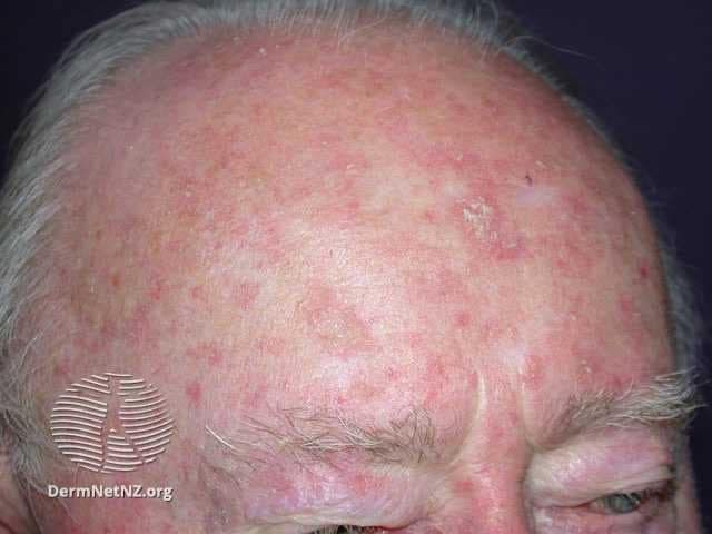
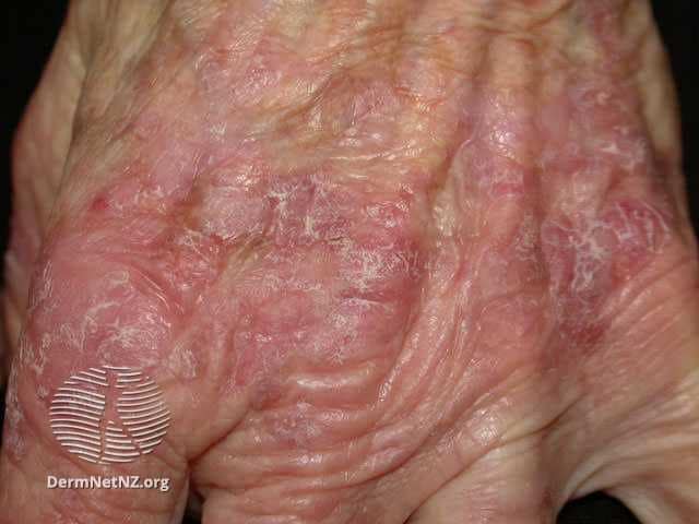
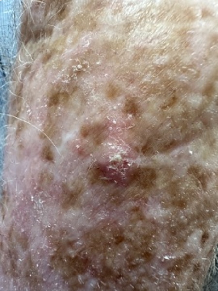
1. Solar Keratosis / Sun Damaged Skin
Solar keratosis, also known as actinic keratosis, is particularly common in Australia due to high UV radiation exposure. These rough, scaly patches on the skin are precursors to squamous cell carcinoma and typically appear in sun-exposed areas like the face and hands.
Treatment Options for Actinic Keratoses
1. Cryotherapy:
Pros: Quick treatment with minimal post-care, suitable for one solitary lesion.
Cons: May require multiple sessions for effectiveness; not suitable for a large field of sun-damaged skin. Risk of hypopigmentation scar.
2. Shave, Curettage, and Electrocautery:
Pros: Immediate removal of the lesion, effective for thick keratoses, suitable for one solitary lesion.
Cons: Some of scarring and requires careful wound care; not suitable for a large field of sun-damaged skin.
3. Topical Treatments:
Two topical cream treatments available.
Pros: Effective at killing precancerous cells. Might be eligible for PBS subsidy. General patient charge $30-35+F17
Cons: Can cause significant skin irritation. Apply to the affected area 2 times a day for 2 to 4 weeks. (Sometimes, not practical). Prolonged treatment duration, may cause local skin reactions.
4. Photodynamic Therapy (PDT):
Pros: Suitable for sun-damaged skin on the face, ears, scalp, and chest; one-day treatment, generally less painful. Downtime of 4-5 days.
Cons: Higher cost compared to other treatments. $600 per region. (Both one side of hands are one region, both one side of forearms is one region). May need 2 treatments depending on your severity.
Prevention of Actinic Keratoses
Apart from practicing sun-smart and attending regular skin checks. (which we are all aware of). Some studies have shown a reduction of Solar Keratosis with:
- Taking Vitamin B3) 500 mg twice.
- Applying prescription Vitamin A.
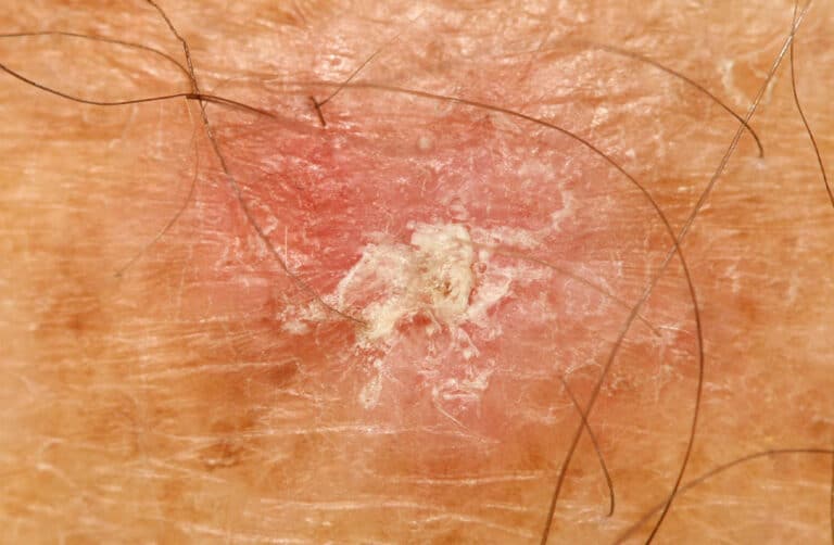
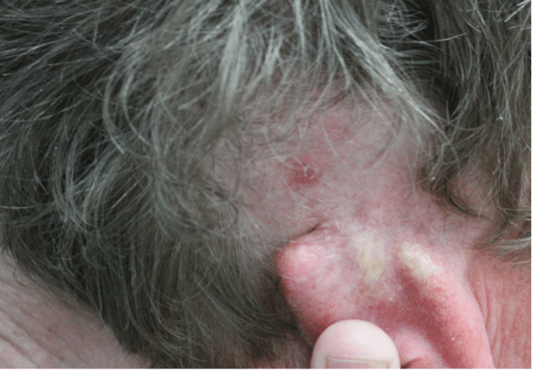
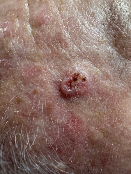
2. Basal Cell Carcinoma (BCC)
Basal Cell Carcinoma (BCC) is the most common form of skin cancer. While BCC is known for its local invasiveness, it rarely metastasises due to its typically slow growth and minimal tendency to spread beyond its original site. However, if left untreated, BCC can cause significant local destruction, potentially eroding through skin and soft tissues to reach underlying bones.
Understanding Basal Cell Carcinoma
BCC arises from mutations in the cells of hair follicles. Consequently, areas without hair follicles—such as the lips, palms, soles, and vulva minora—are unlikely to develop BCC.
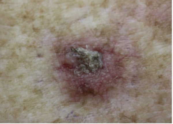
Symptoms: BCC typically appears as a skin-coloured, pink, or pigmented nodule or plaque with a pearly edge and can ulcerate. Different types include nodular, superficial, morphoeic, and the more aggressive basosquamous carcinoma.
Treatment Necessity
Despite its low risk for metastasis, BCC requires treatment to prevent extensive local damage. If you have an upcoming holiday or important event. You can have up to 6 months to schedule your excision. Treatment options include –
- Surgical Excision: Often the first choice, providing clear margins to ensure complete removal.
- Mohs Surgery: Ideal for facial and other complex areas, preserving healthy tissue while thoroughly removing cancer cells. We will involve your care with a Dermatologist who specialises in Mohs Surgery.
- Less Invasive Treatments: Cryotherapy, photodynamic therapy, and topical treatments are options for less aggressive or superficial BCCs.
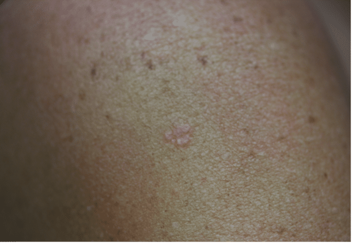
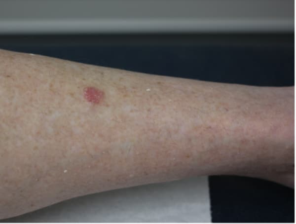
3. Squamous Cell Carcinoma (SCC)
Squamous Cell Carcinoma (SCC) is a type of skin cancer that can develop from precursors like actinic keratosis, a result of prolonged UV exposure. Without intervention, actinic keratosis can progress to intraepidermal carcinoma (IEC) and then to invasive SCC, which may penetrate deeper skin layers and potentially metastasise.
This progression highlights the importance of early detection and preventive treatment.
Preventative Treatment
Preventive measures are crucial in managing the risk of SCC. While acknowledging the benefits of Sunlight for vitamin D synthesis and mental health, it is essential to balance this with protective strategies to minimise sun damage.
The Importance of Early Treatment
Addressing skin changes promptly when they are less severe—not only saves skin but also lives. Treatments for early stages, like cryotherapy for actinic keratosis, topical medications for IEC, PDT field treatments are generally simple and highly effective. These early interventions prevent the progression to invasive SCC, thereby reducing the risk of metastasis and more complicated treatments required at later stages.
Long-Term Monitoring
Once treated for any stage of SCC progression, ongoing monitoring is essential. Regular follow-ups of 6monthly visits are recommended as this help catch any new changes early and maintain healthy skin over the long term.
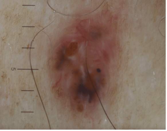
4. Melanoma
Understanding Melanoma: Australia’s National Cancer
Australia has the highest melanoma rates worldwide, often called ‘Australia’s national cancer.’ It’s crucial to recognise its prevalence and severity because of its potential for early detection and successful treatment. Melanoma can spread to many body parts, commonly affecting the lymph nodes, lungs, liver, bones, and brain. Early detection is key to managing its spread effectively.
Key Statistics
- Annual Diagnoses: Approximately 16,800 Australians are diagnosed each year.
- Mortality Rate: Around 1,300 Australians die annually from melanoma.
- Most common cancer in Australians aged 20 to 39.
- Second most common in men, after prostate cancer.
- Third most common in women, after breast and colorectal cancer.
Prevention and Detection
Early detection can lead to a 90% cure rate through surgical intervention.
Significant progress has been made in the last decade, with the 5-year survival rate for advanced melanoma increasing from less than 10% to over 50%.
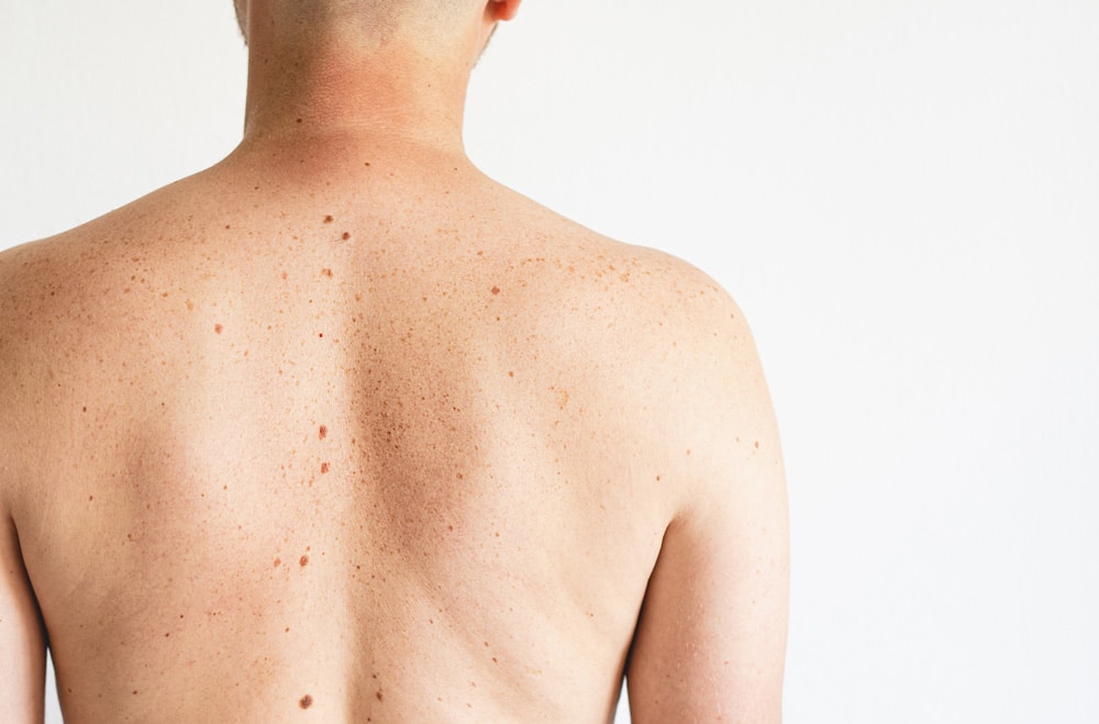
How to Check Your Skin
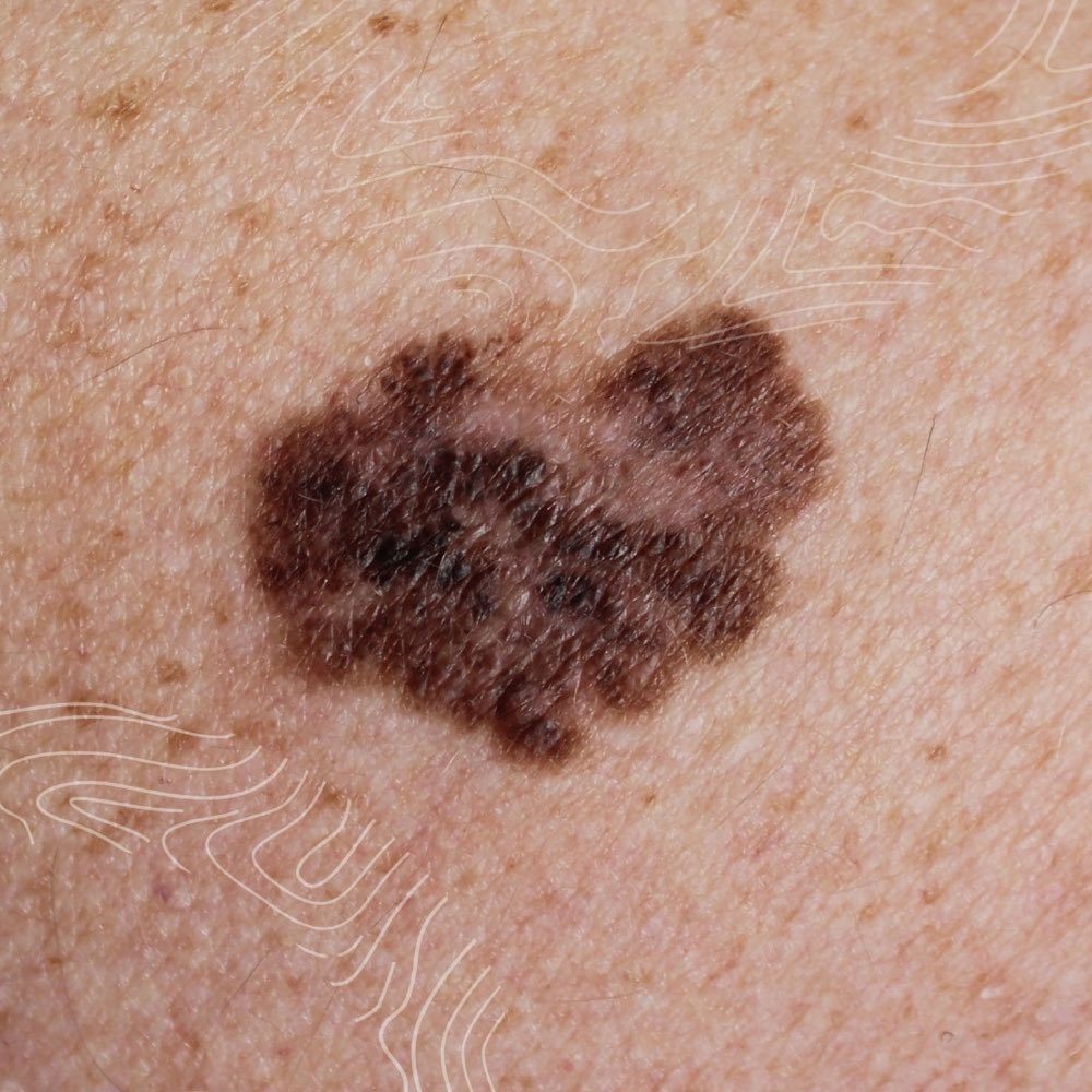
ABCDE Method of Identifying Melanoma
A for Asymmetry: One half of the spot does not match the other.
B for Border: Edges are irregular, ragged, or blurred.
C for Colour: Varies within the same spot.
D for Diameter: Larger than 6mm or growing.
E for Evolving: Any change in size, shape, colour, or symptoms such as itching or bleeding.
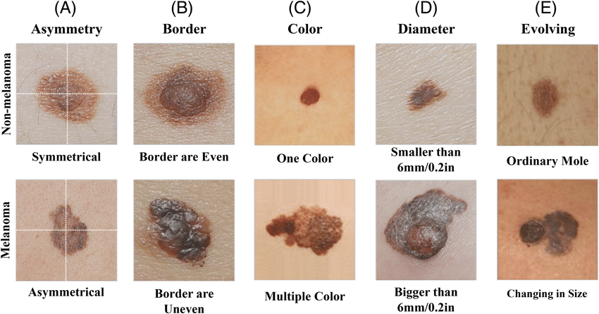 Image source: https://www.researchgate.net/figure/Illustration-of-ABCDE-criteria-for-skin-cancer-detection_fig2_359338536
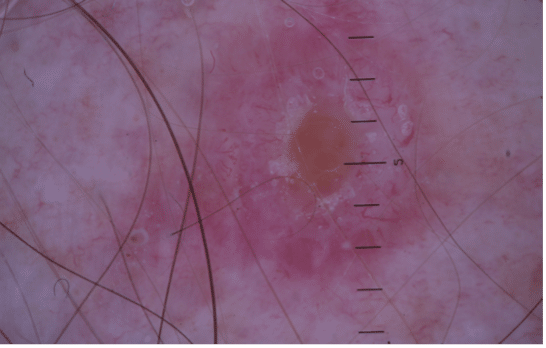
If you notice any changes or new spots on your skin, don’t hesitate to book a quick spot check. Dr. Tina Fang and Dr. Jack Fu are committed to addressing your concerns promptly. Even if you suspect it might be nothing, it’s always best to have it examined. At our clinic, we use Dermatoscopy to provide a thorough assessment, ensuring that even the smallest changes are carefully evaluated. It’s always better to be safe and have any changes checked by our experts.