When discussing skin cancer, most people think of melanoma or basal cell carcinoma. However, there is another category of skin malignancies that, whilst less commonly discussed, represents an important clinical concern and an excellent opportunity for early intervention. Intraepidermal carcinoma, also known as squamous cell carcinoma in situ or Bowen’s disease, is a precancerous lesion that, if left untreated, can progress to invasive squamous cell carcinoma. At ISO Skin Cancer & Laser Clinic, we recognise the critical importance of early detection and treatment of these lesions, and as a candidate for the Master of Skin Cancer Medicine at the University of Queensland, I, Dr. Tina Fang, have devoted significant research to understanding the optimal treatment strategies for intraepidermal carcinoma.
This blog post will define intraepidermal carcinoma and Bowen’s disease, explain the risk of progression to invasive cancer, and discuss why PDT is an ideal first line treatment for these conditions.
Defining Intraepidermal Carcinoma and Bowen’s Disease
Intraepidermal carcinoma is a malignant lesion that is confined to the epidermis (the outermost layer of the skin) without invasion into the dermis (the layer of skin beneath the epidermis). When this condition occurs on the genitals, it is referred to as erythroplasia of Queyrat (in men) or Bowen’s disease of the genitals (in women). When it occurs on non genital skin, it is simply referred to as Bowen’s disease or squamous cell carcinoma in situ.
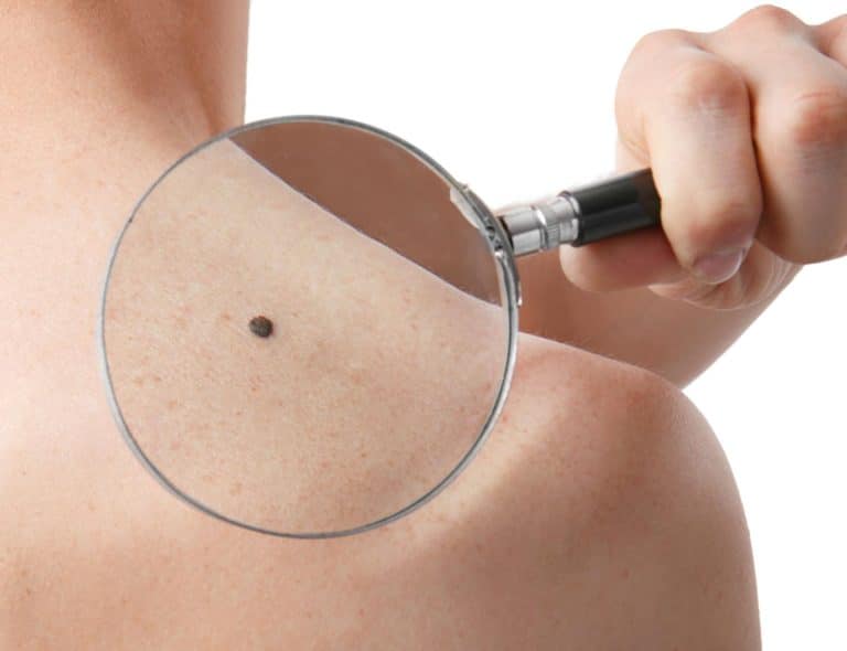
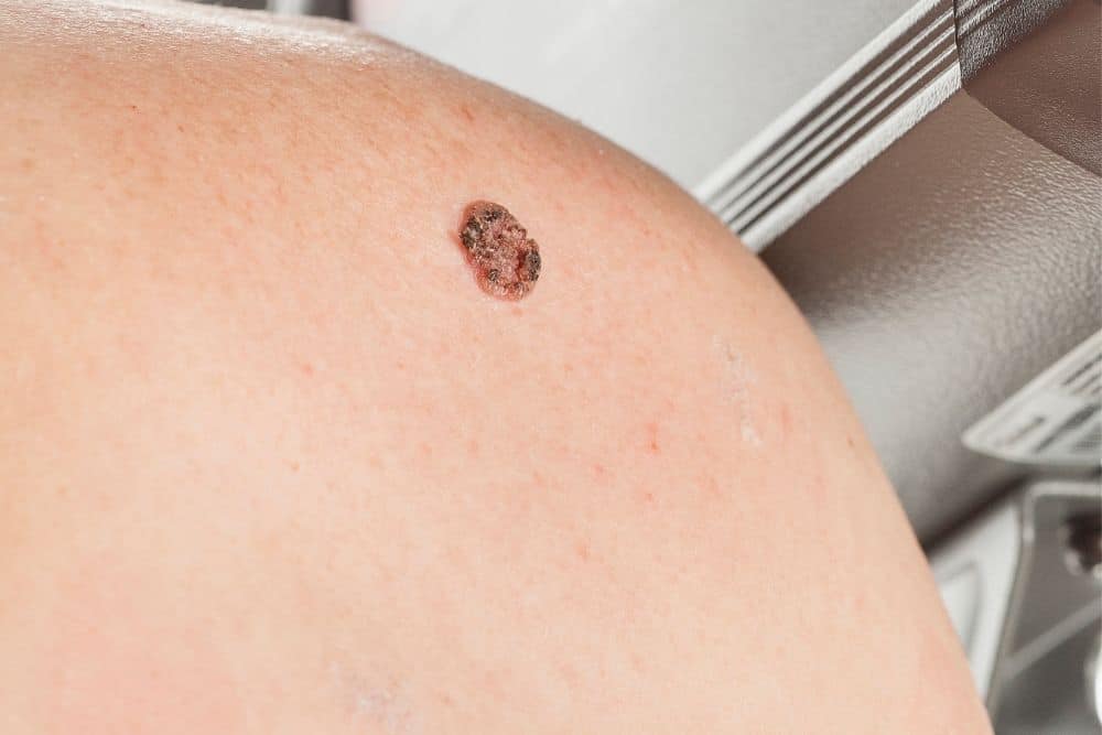
Bowen’s disease typically appears as a well demarcated, erythematous (red), scaly patch or plaque on the skin. The lesion may have an irregular border and can vary in size from a few millimetres to several centimetres. The surface may be crusted or bleeding, and the patient may report itching or tenderness. These lesions most commonly occur on sun exposed areas of the body, such as the face, scalp, hands, and forearms, although they can occur anywhere on the skin.
Risk of Progression to Invasive Cancer
The critical distinction between intraepidermal carcinoma and invasive squamous cell carcinoma is the presence or absence of invasion into the dermis. Intraepidermal carcinoma, by definition, does not invade the dermis. However, if left untreated, intraepidermal carcinoma can progress to invasive squamous cell carcinoma, which carries a significantly higher risk of metastasis and mortality.
Studies have shown that approximately 3% to 5% of untreated Bowen’s disease will progress to invasive squamous cell carcinoma. Whilst this percentage may seem relatively low, it represents a significant risk, particularly when one considers that a patient may have multiple lesions or may develop new lesions over time. Additionally, the risk of progression is higher in immunocompromised patients, such as those with HIV or those taking immunosuppressive medications.
Therefore, prompt treatment of intraepidermal carcinoma is essential to prevent progression to invasive disease and to reduce the overall burden of malignancy in the skin.
Why PDT is Ideal for These Conditions
Photodynamic Therapy is an ideal treatment for intraepidermal carcinoma and Bowen’s disease for several reasons. First, PDT is highly selective for abnormal cells, preferentially targeting the rapidly dividing cells of the malignancy whilst largely sparing the surrounding healthy tissue. This selectivity is particularly important for lesions located in cosmetically sensitive areas or on thin skinned areas where surgical excision might result in significant scarring or functional impairment.
Second, PDT can be used to treat multiple lesions in a single session, making it an efficient treatment option for patients with extensive disease or field cancerisation. Third, PDT is non invasive and does not require anaesthesia, making it a well tolerated treatment that can be performed in an outpatient setting. Finally, PDT has been shown to have excellent cure rates for intraepidermal carcinoma, with clinical clearance rates exceeding 90% in most studies.
Histopathologic Evidence of Treatment Success
The effectiveness of PDT for intraepidermal carcinoma is not only evident from clinical observation but is also confirmed by histopathologic examination. A study published in Dermatologic Surgery evaluated the use of PDT with intense pulsed light for actinic keratosis and found that PDT caused the disappearance of abnormal keratinocytes from the epidermis . Importantly, the study also noted that some lesions showed atypical cells in the follicular infundibulum, suggesting that in some cases, abnormal cells may persist in deeper structures and may require additional treatment or close follow up.
This histopathologic evidence underscores the importance of careful patient selection, appropriate treatment parameters, and diligent follow up to ensure complete resolution of the lesion and to detect any recurrence at the earliest opportunity.
Multiple Treatment Protocols and Their Effectiveness
There are several treatment protocols for PDT that can be used for intraepidermal carcinoma, and the choice of protocol depends on various factors including the size and location of the lesion, the patient’s skin type, and the patient’s tolerance for treatment related side effects.
The most commonly used protocol involves the application of a 20% 5 ALA cream, which is left on the skin for approximately 2 hours before light activation. This protocol has been shown to be highly effective, with clinical clearance rates of 85% to 95% in most studies. An alternative protocol involves the use of methyl aminolevulinate (MAL), which has a shorter incubation time of approximately 30 minutes to 1 hour, making it a more convenient option for some patients.
Laser assisted PDT, which combines fractional laser pretreatment with PDT, has been shown to achieve even higher cure rates, often exceeding 95%. This approach is particularly useful for thicker lesions or lesions that have not responded adequately to standard PDT.
| PDT Protocol | 5 ALA Concentration | Incubation Time | Light Source | Effectiveness |
| Standard PDT | 20% cream | 2 hours | LED or laser | 85 95% |
| MAL PDT | 16% ointment | 30 60 minutes | LED or laser | 80 90% |
| Laser Assisted PDT | 20% cream or MAL | 60 90 minutes | Intense pulsed light | 95%+ |
Monitoring for Recurrence
Following PDT treatment of intraepidermal carcinoma, it is essential to maintain close follow up to monitor for recurrence. Most recurrences occur within the first 1 to 2 years after treatment, so frequent clinical examinations during this period are important. Your cosmetic physician may recommend follow up visits at 1 month, 3 months, 6 months, and 12 months after treatment, and then annually thereafter.
If recurrence is suspected, a skin biopsy may be performed to confirm the diagnosis. If recurrence is confirmed, additional PDT treatment can usually be performed, or alternative treatment modalities such as surgical excision or laser ablation may be considered.
Preventive Strategies for High Risk Patients
Patients who have had intraepidermal carcinoma are at increased risk of developing additional skin cancers in the future. This is particularly true for patients with extensive photodamage or those with a history of significant sun exposure. Therefore, it is crucial to adopt comprehensive preventive strategies following treatment.
These strategies include daily use of a broad spectrum sunscreen with an SPF of 50+, wearing protective clothing such as a wide brimmed hat and sunglasses, seeking shade during peak UV hours, and avoiding deliberate sun exposure. Additionally, regular skin self examination and professional skin checks are essential for early detection of any new lesions.
Some patients may benefit from prophylactic treatment with topical retinoids or other chemoprevention agents, which have been shown to reduce the risk of developing new actinic keratoses and other precancerous lesions. Your cosmetic physician can advise you on whether such preventive measures are appropriate for your individual situation.
Treatment Considerations and Patient Selection
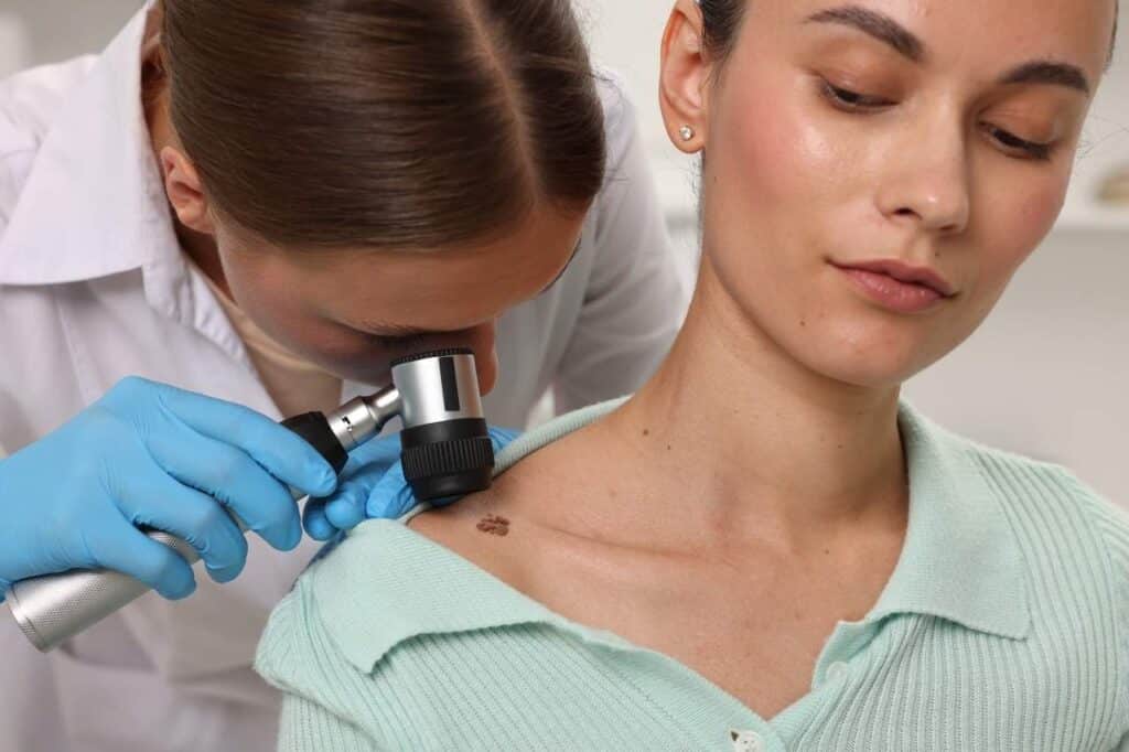
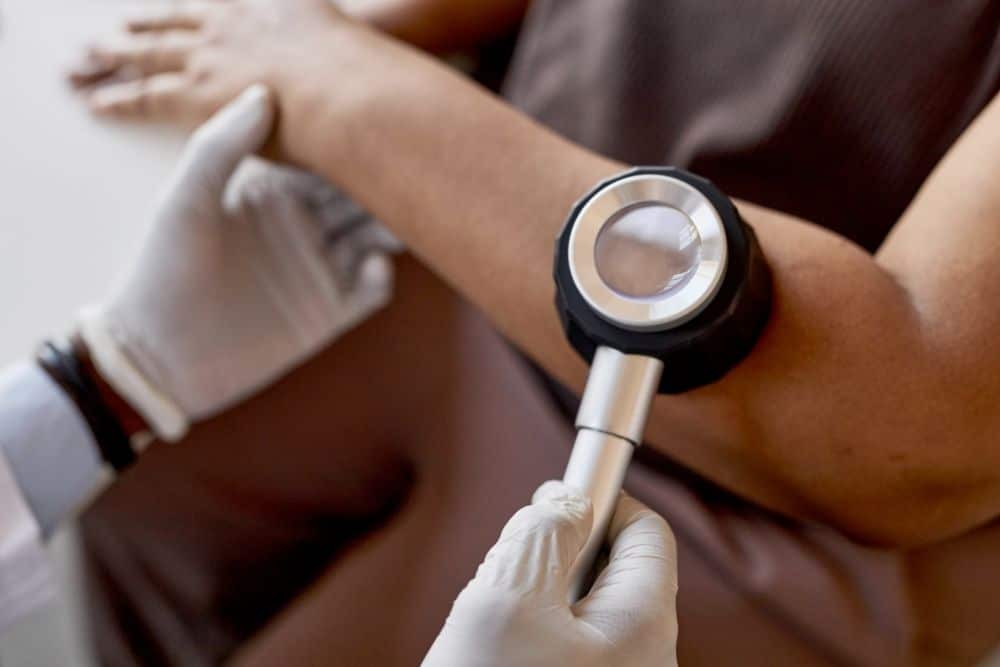
Not all patients with intraepidermal carcinoma are suitable candidates for PDT. Certain factors, such as the size and location of the lesion, the patient’s skin type, the patient’s immune status, and the patient’s ability to comply with post treatment care instructions, must be considered. A thorough consultation with a qualified cosmetic physician is essential to determine if PDT is the appropriate treatment for your individual situation.
During your consultation, your cosmetic physician will examine the lesion, perform any necessary diagnostic tests (such as a skin biopsy to confirm the diagnosis), and discuss the available treatment options. Together, you will develop a personalised treatment plan that addresses your specific needs and maximises the likelihood of achieving your desired outcomes.
Conclusion
Intraepidermal carcinoma and Bowen’s disease represent important clinical entities that require prompt and effective treatment to prevent progression to invasive squamous cell carcinoma. PDT, with its high cure rates, minimal scarring, and excellent cosmetic outcomes, represents an ideal first line treatment for these conditions. For patients concerned about their skin’s health or those who have been diagnosed with intraepidermal carcinoma, PDT offers an evidence based, non invasive treatment option that can effectively eliminate the malignancy and reduce the risk of future skin cancer development.
At ISO Skin Cancer & Laser Clinic, we are committed to offering our patients the latest, evidence based treatments for skin cancer. If you have been diagnosed with intraepidermal carcinoma or Bowen’s disease, or if you are concerned about your skin’s health, we encourage you to schedule a consultation with one of our experienced cosmetic physicians to discuss your treatment options.
References
- Kim, H. S., Yoo, J. Y., Cho, K. H., Kwon, O. S., & Moon, S. E. (2005). Topical Photodynamic Therapy Using Intense Pulsed Light for Treatment of Actinic Keratosis: Clinical and Histopathologic Evaluation. Dermatologic Surgery, 31(1), 33 37.
- Sullivan, J. R., & Sharpe, P. D. (2021). Photodynamic Therapy for Superficial Sun Damage. Opinions and Progress in Cosmetic Dermatology, 1(3), 36 40.